


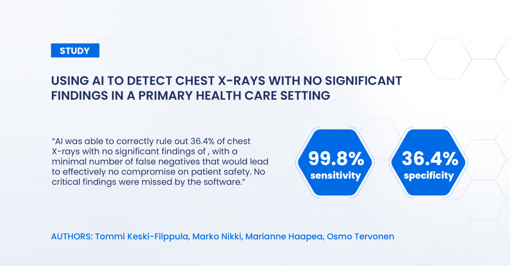
Following the CE certification of the first autonomous AI medical imaging application ChestLink, researchers in Finland released a preprint of a study on the application performance on real-world clinical data. In the study, the application analyzed 10.000 chest X-rays of Finnish primary care patients. ChestLink software was able to identify normal studies with 99.8% sensitivity and and specificity of 36.4% with a minimal number of false negatives that would lead to effectively no compromise on patient safety and no critical findings missed by the software.
After the exclusion of cases not meeting the study criteria, 9579 retrospective cases were analyzed by Oxipit ChestLink. Of these cases, 4451 were considered normal in the original radiologist report and 4644 after the consensus reading. The number of cases correctly found nonsignificant by AI was 1692 (17.7% of all studies and 36.4% of studies with no significant findings).
After the consensus read, there were nine confirmed false-negative studies. These studies included four cases of slightly enlarged heart size, four cases of slightly increased pulmonary opacification and one case with a small unilateral pleural effusion.
Yet according to the researchers, the potential effect of these misdiagnoses on patient safety is negligible and none of them would lead to immediate threat to the patient. The research paper states that “a slightly enlarged heart size is usually not an acute finding, except in the rare case of pericardial fluid, and does not require immediate treatment. A small amount of pleural fluid has little clinical relevance. The four cases with a slightly increased pulmonary opacification might indicate mild bronchopneumonia, but the finding is controversial, and interobserver variability is high among radiologists in reporting this finding”.
The researchers conclude that AI can reliably remove 36.4% of normal chest X-rays from a primary health care population data set with a minimal number of false negatives, leading to effectively no compromise on patient safety. No critical findings, such as lung masses, pneumothoraces or lobar consolidations were missed by the software.
In the opinion of researchers, using AI to rule out normal studies could provide a way to optimize the use of resources in healthcare. With the increasing volumes of CT and MRI, the radiologist workforce could thus be directed to more complicated studies.
The study was conducted by researchers at Oulu University Hospital: Tommi Keski-Filppula, Marko Nikki, Marianne Haapea and Osmo Tervonen, with Oxipit providing ChestLink software for CXR analysis. The authors state that this work has been conducted in an impartial manner and not received any funding.
About Oxipit

Oxipit develops AI applications for diagnostic medical imaging. With a team of award-winning data scientists and medical doctors, the company aims to introduce innovative artificial intelligence breakthroughs to everyday clinical practice.