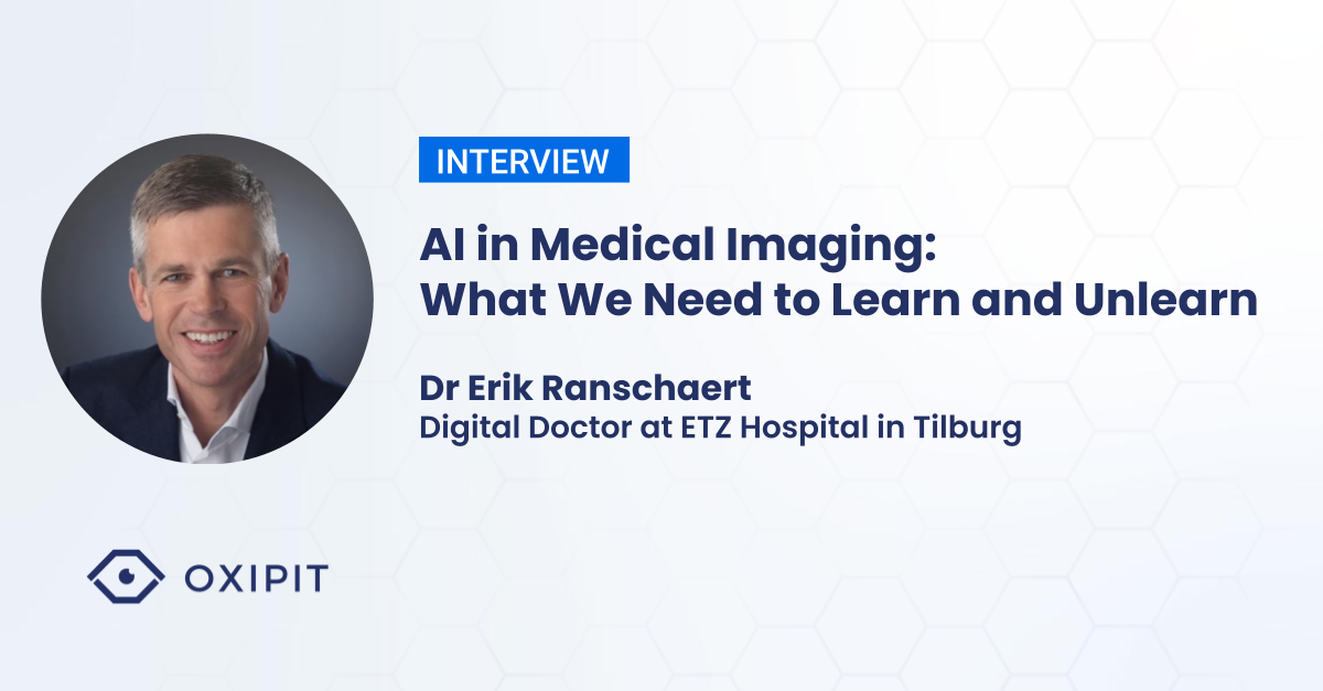
Dr Erik Ranschaert is one of the key AI medical imaging experts in Europe. He is Digital Doctor at ETZ Hospital in Tilburg, responsible for developing a hospital-wide AI infrastructure.
In this interview with Oxipit, Erik shares his views on AI tool adoption, future of radiology and why we should learn and unlearn.
In your opinion, have AI medical imaging tools already made an impact on day-to-day radiology practice?
The advent of AI medical imaging tools challenges medical practitioners to learn new and unlearn traditional things.
Chest X-rays are the staple of medical imaging. Together with musculoskeletal imaging, they are the most common diagnostic procedure.
At our hospital around 40-50% of chest X-rays contain no abnormalities. If AI can cover and report on those cases – even if not all – this is already a huge boost to productivity. While our department is not short staffed, this is not the case in many European countries or the developing world, where due to capacity problems chest X-rays cannot even be reported by radiologists.
Furthermore AI helps to move radiology from morphological to a more structured, functional reporting. This lays the foundation for a more data-driven diagnostic and medical care environment.
As we deploy AI tools in medical institutions, we are constantly discovering new facets of the traditional framework – for instance, in radiology resident training or empowering radiographers to verify the final ‘no-findings’ reports. There is a lot of potential we are yet to uncover.
As for unlearning, AI tools push radiologists to unlearn that they are working on their own. AI is a well-informed assistant operating in the background. After a report by a radiologist is filed, if AI finds a mismatch with its own findings, it invites the medical special to take a second, more detailed look.
Even if AI is not ‘correct’ in all of these cases, we welcome the fact that the tool could bring our attention to something we might not have noticed ourselves. This AI ‘prompt’ already adds value to the diagnostic process.
For a few years now medical imaging AI tools have been a part of the radiology landscape. What is the radiologist’s opinion on AI and how it has changed in the past years?
Recently we have conducted a global radiologist survey on AI technologies. After some initial distrust, radiologists do not see these tools as a threat anymore. They grew to understand that they are made for a specific task and that they can improve reporting quality, aid decision making or increase productivity.
The most skepticism is reported from the specialists who have the least information or experience with AI.
Education is key. Even when we approach reporting autonomy, it is crucial to understand that humans are still very much in the loop – in terms of auditing, traceability or overruling the AI decisions.
The same goes with wider understanding in the general public – hospital patients. It should be made clear that radiology specialists and their virtual AI assistants are working on their case as a team – to reduce the chance of errors and arrive at a more precise diagnosis.
What benefits does early AI adoption bring to the first-mover medical institutions?
In certain environments, such as teleradiology which is commercially competitive, early adoption of AI tools can serve as a competitive advantage.
As for the case of most hospitals, it is about being ahead of the field and using the latest tools and methods available in medical practice.
It certainly helps to attract talent to the hospital’s radiology department, develop new methods of practitioner training and prepare the next generation of radiology specialists.
This is not limited to AI in medical imaging. Overall, the adoption of AI tools in all fields of medical practice help to position the hospital as a cutting-edge medical institution.
Considering the adoption of new methods and technologies, how would a typical radiology department look and operate in 20 years time?
In the past few decades radiology has moved into the digital realm. We now have more information and are learning to do more with this information. We are also shifting from morphological, to a structured, functional approach to reporting.
This means that radiology will become more like data analysis. As radiologists, we will need to interpret and manage this data – with the assistance of technological tools.
Imaging data integration into a holistic healthcare infrastructure will serve as a part of the foundation of precision medicine – where we can personally approach the treatment of a specific patient, resulting in reduced costs and better patient outcomes.


