Last April Oxipit took the medical imaging world by storm. The medical imaging company received a CE Class IIb certificate for the first autonomous AI imaging application ChestLink. The application produces final chest x-ray study reports where it is highly confident that the study features no abnormalities – without any involvement from a radiologist. To this day ChestLink remains the only certified AI radiology application capable of autonomous performance.
“Everyone was expecting AI medical imaging applications to operate autonomously – five, ten or even more years in the future. Nobody was expecting for this to happen in 2022. Furthermore, this innovation did not come from a big tech outfit somewhere in Silicon valley. It happened in Vilnius, European Union, made possible by a dedicated team of medical and data science specialists”, – says CEO of Oxipit Gediminas Peksys.
Vanguard of Artificial Intelligence
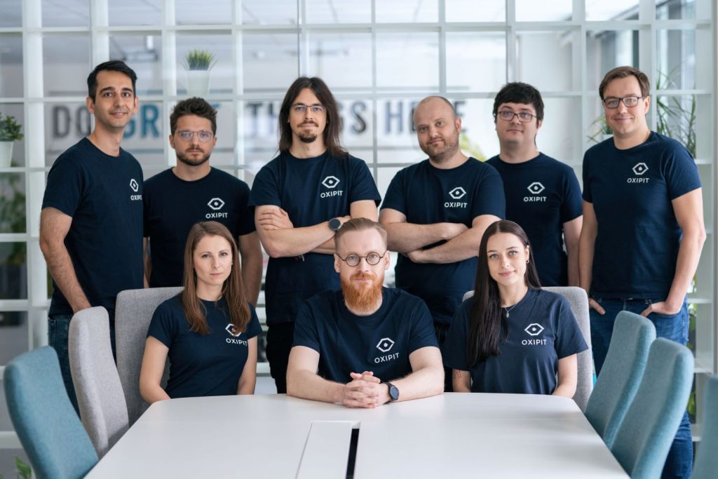
A Cambridge graduate, a Kaggle grandmaster, an astrophysicist and a radiologist walk into a bar… That could be the setup for a good joke. Yet it is not far from the truth in the story of how Oxipit was founded.
“More than 5 years ago four of us met at an AI hackathon. Everyone from the team had accomplishments in their respective scientific or professional fields. Some of us had previous experience of developing world class AI applications for object recognition and image analysis – yet as employees, not company founders. Everyone in the team had ambitions to build something on their own”, – remembers Gediminas Peksys.
In the hackathon the team focused on the global shortage of radiologists. Over the course of 24 hours the prototype for Oxipit ChestEye was born.
“From a data science perspective, the medical imaging field was a good choice to utilize our advantage in image analysis. It also presented a fundamental global healthcare problem, where AI could make an impact. The global population is aging. The demand for medical imaging increases every year. We are not training radiologists fast enough. In developing countries, the rate of 1 radiologist per millions of patients is the sad norm. AI could bridge this gap and significantly improve patient care sooner rather than later”, – says Gediminas Peksys.
Moving past CAD
Oxipit ChestEye received CE certification in 2019. The AI CAD application produced preliminary reports for 75 chest x-ray findings. ChestEye became the backbone of next generation Oxipit application development.
“From the get go we were thinking on how a radiology department in the future would look like. Due to radiologist subjectivity – and they tend to disagree quite often – AI CAD applications only provide limited gains in real world medical practice”, – says Chief Medical Officer at Oxipit Naglis Ramanauskas.
According to Dr Ramanauskas, who was and still is a practicing radiologist, Oxipit products should evolve into an integral part of radiology workflows, reducing radiologist workflow and improving diagnostic performance.
“Cutting edge AI algorithm performance is only the backbone for a great product. It is about defining the usage scenarios and workflows. No extra screens, no extra button taps, no distractions, objective tangible value for the radiologist – only then AI becomes an utility that medical specialists actually want to use”, – says Naglis Ramanauskas.
Aiming for autonomous AI imaging
Darius Barusauskas is the Chief Data Science Officer at Oxipit and a 4x Kaggle Grandmaster. Akin to ATP rankings for tennis, Kaggle ranks data scientists on their performance in data science competitions. At one time Darius held a 4th ranking overall from hundreds of thousands of experts.
“What attracted me to the medical imaging field is the complexity of machine learning problems and the value it can bring to patient care if these issues are solved”, – tells Darius Barusauskas.
He regarded autonomous AI imaging algorithm development as one of his personal and professional ambitions. From the inception of Oxipit, it took 4 years for this dream to start to become true.
When working on ChestLink, the team selected no-abnormality reports as the first step for autonomous AI imaging application. The application automates the scope of radiologist invariant chest X-rays, which are of normal appearance to any given radiologist. These studies can be autonomously reported by the application and removed from the radiologist workflow. In case there is a slightest suspicion that an X-ray may feature an abnormality, the application leaves the reporting of the study to the hospital radiologist.
More than 99% sensitivity
ChestLink was trained on a 500K+ training image dataset.
In impartial clinical validation trials the application consistently performed at 99%+ sensitivity rate.
A research study published in the Radiology Journal in March highlighted the sensitivity of the application.
“The most surprising finding was just how sensitive this AI tool was for all kinds of chest disease. In fact, we could not find a single chest X-ray in our database where the algorithm made a major mistake. Furthermore, the AI tool had a sensitivity overall better than the clinical board-certified radiologists”, – said the study co-author Louis Lind Plesner, MD, from the Department of Radiology at the Herlev and Gentofte Hospital in Copenhagen, Denmark.
In this study ChestLink could have autonomously reported on 28% of all normal studies.
In another ChestLink validation study at the Oulu University Hospital, the researchers concluded that the autonomous AI imaging application can reliably remove 36.4% of normal chest X-rays from the reporting workflow with a minimal number of false negatives, leading to effectively no compromise on patient safety. In the study ChestLink software was able to identify normal studies with 99.8% sensitivity.
“Our initial trials, current deployments and third-party validation has consistently shown that ChestLink can operate at or exceeding the sensitivity level of human radiologists, without any compromises on patient safety”, – added Gediminas Peksys.
Preventing diagnostic mistakes
Radiologists operate in a cognitively intense real time environment. They work solo. Mistakes happen to the best of us. Yet radiology lacks effective frameworks for quality control.
“Double reading audits happen after the fact. We wanted to create a useful tool to catch diagnostic mistakes in real-time, so the treatment action can be augmented before, for instance, a patient is discharged from the hospital”, – tells Naglis Ramanauskas.
Oxipit Quality is an AI-powered second reader application. It operates as an always-on radiologist assistant. The AI application annalizes radiological images and corresponding radiologist reports.
If the application detects a clinically impactful finding, which is absent from the radiologist report, it sends an automated notification to the radiologist to take another look.
“We call it a ‘safety-net’. The application already proved instrumental in improving subtle pulmonary nodule detection, helping to detect lung at an earlier stage”, – says Naglis Ramanauskas.
From clinical studies across the board, the rate of significant missed findings varies from 0.2 to 1% depending on the medical institution.
“While this is a relatively low percentage, it still represents a significant number of potential errors that could be caught with the use of an AI-based tool. If you look at the scale of the operation of our institution, in which we do over 300,000 exams a year, that means 3,000 patients who would have a change in their treatment outcomes. Even such small numbers could change the lives of so many families”, – says Chief Medical Officer at CARPL.ai Dr Vasanth Venugopal, who recently led a validation study of Oxipit Quality.
Urgent Need, Careful Use
NHS England recognises the need to support AI adoption against a backdrop of 7 million chest xrays performed annually in England alone, and with a shortage of 1,669 consultant radiologist and has launched the AI Diagnostics Fund; a unique opportunity to address some of the biggest challenges faced by Radiology Departments throughout NHS England.
Healthcare IT veteran and Oxipit Sales Manager Peter Corscadden recognises the need to support NHS customers contemplating autonomous reporting with the option of gradual introduction of autonomy into the CXR workflow. “Luckily the AIDF allows us to support NHS England customers with the suite of Oxipit CXR solutions and we can configure each deployment according to local preference in terms of autonomy. Our partnership with an NHS Foundation Trust has already shown us the value of a collaborative approach”.
Leapfrog technologies
With a shortage of consulting radiologists and growing reporting backlogs, the initiative calls for quick results from forthcoming diagnostic AI application deployments.
“In the context of available AI solutions, Oxipit products are leapfrog technologies – enabling healthcare providers to skip the limitations of typical CAD applications and aim for tangible, swift clinical impact”, – summarizes Gediminas Peksys.
Learn more about Oxipit AIDF offering at https://oxipit.ai/nhs-aidf-application/


